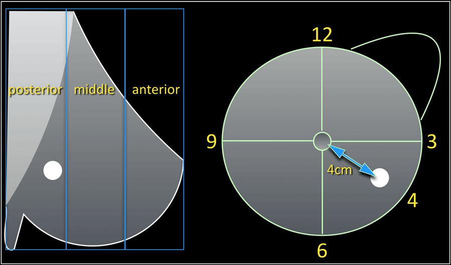
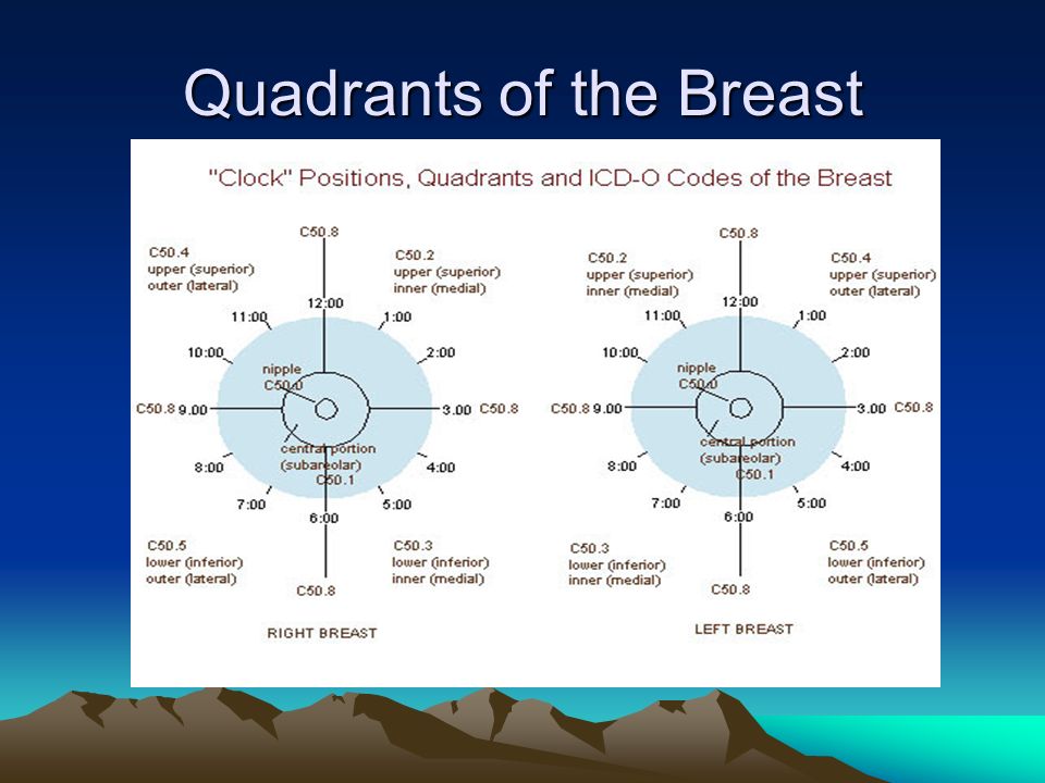
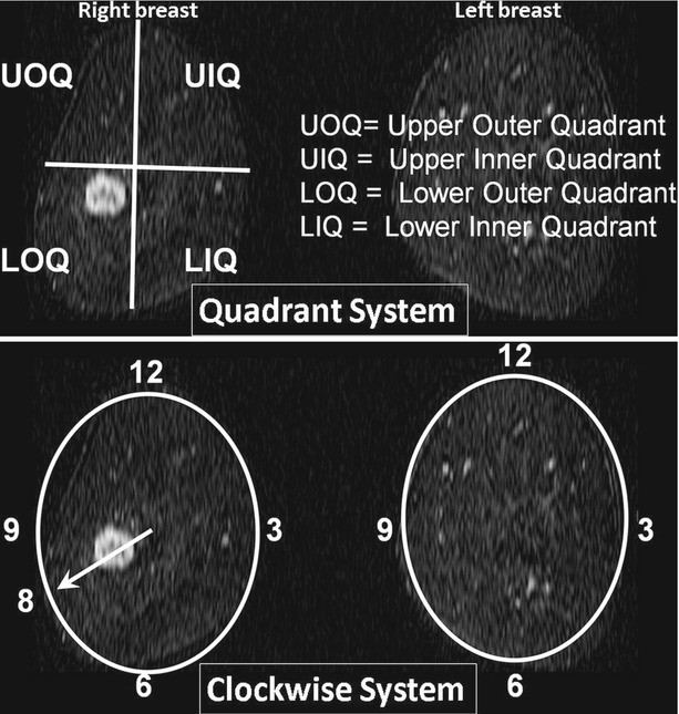
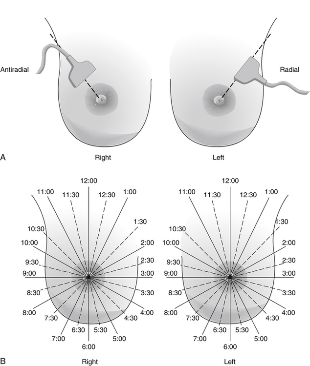
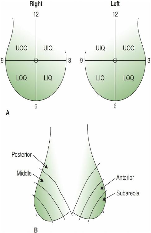
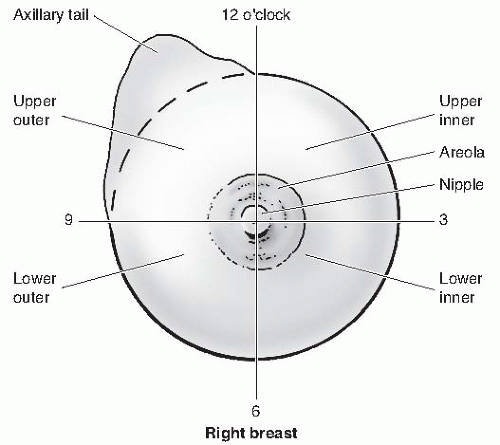
0 #4 Breast quadrants According to SuperCodercom "The correct codes for the 12 o'clock position breast mass are N6322 and N6321 or N6311 and N6312 as per laterality mentioned in document" Another post from from terrilynnlogan@gmailcom states "There was a WPS call back on Jan 10th where this issue was addressedCode C509 (Breast, NOS) in this situation Code the primary site to C508 when O'Clock Positions and Codes Quadrants of Breasts 2 11 12 1 1 10 9 8 7 7 6 5 4 3 2 11 12 10 6 5 3 RIGHT BREAST LEF T BREAST UOQ UIQ UIQ UOQ LOQ LIQ LIQ LOQ C504Position, the upper limb of the affected side was lifted up into the same position as the intraoperative position, and the breast and axilla of the affected side was fully exposed An intradermal injection of 1 mL UCA at 3 and 6 o 'clock and a subcutaneous injection of 1 mL UCA at 9 and 12 o 'clock of the mammary areola region

The Radiology Assistant Bi Rads For Mammography And Ultrasound 13
Where is 10 o'clock position on breast
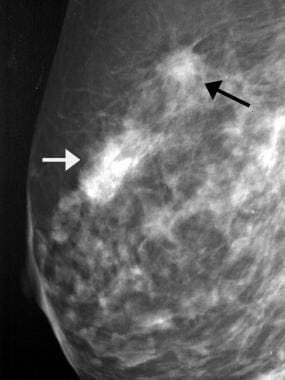
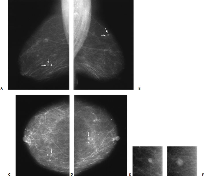
Where is 10 o'clock position on breast-Multicentric breast cancer in a 63yearold woman Right mediolateral oblique (a) and right exaggerated craniocaudal lateral (b) screening mammograms show a prominent area of architectural distortion at the 10 o'clock position (solid arrow) Note also the two small, indistinct masses in the axillary tail (arrowheads) and the linearly arranged microcalcifications at the 7Code C509 (Breast, NOS) in this situation Code the primary site to C508 when • there is a single tumor in two or more subsites and the subsite in which the tumor originated is unknown • there is a single tumor located at the 12, 3, 6, or 9 o'clock position on the breast


1
There is a 7mm hypoechoic nodule with shadowing at the 10 o'clock position of the right breast The biopsy sampling of the ill defined opacity in the upper outer right breast on mammography Also, biopsy of the 7mm hypoechoic solid nodule at the 10 o'clock position in the right breast, only seen with sonography Birads category 4Code C509 (Breast, NOS) in this situation O'Clock Positions and Codes Quadrants of Breasts 2 11 12 1 1 10 9 8 7 6 5 7 4 3 2 11 12 10 6 5 4 3 RIGHT BREAST LEF T BREAST UOQ UIQ UIQ UOQ LOQ LIQ LIQ LOQ C504 C502 C502 C504 C505 C503 C505 C503 C500 C501Gravity A lesion located in the upper outer quadrant of the right breast is located in the 1 5o'clock position 2 2o'clock position 3 10o'clock position 4 7oc'clock position Click card to see definition 👆 Tap card to see definition 👆 3 10o'clock position
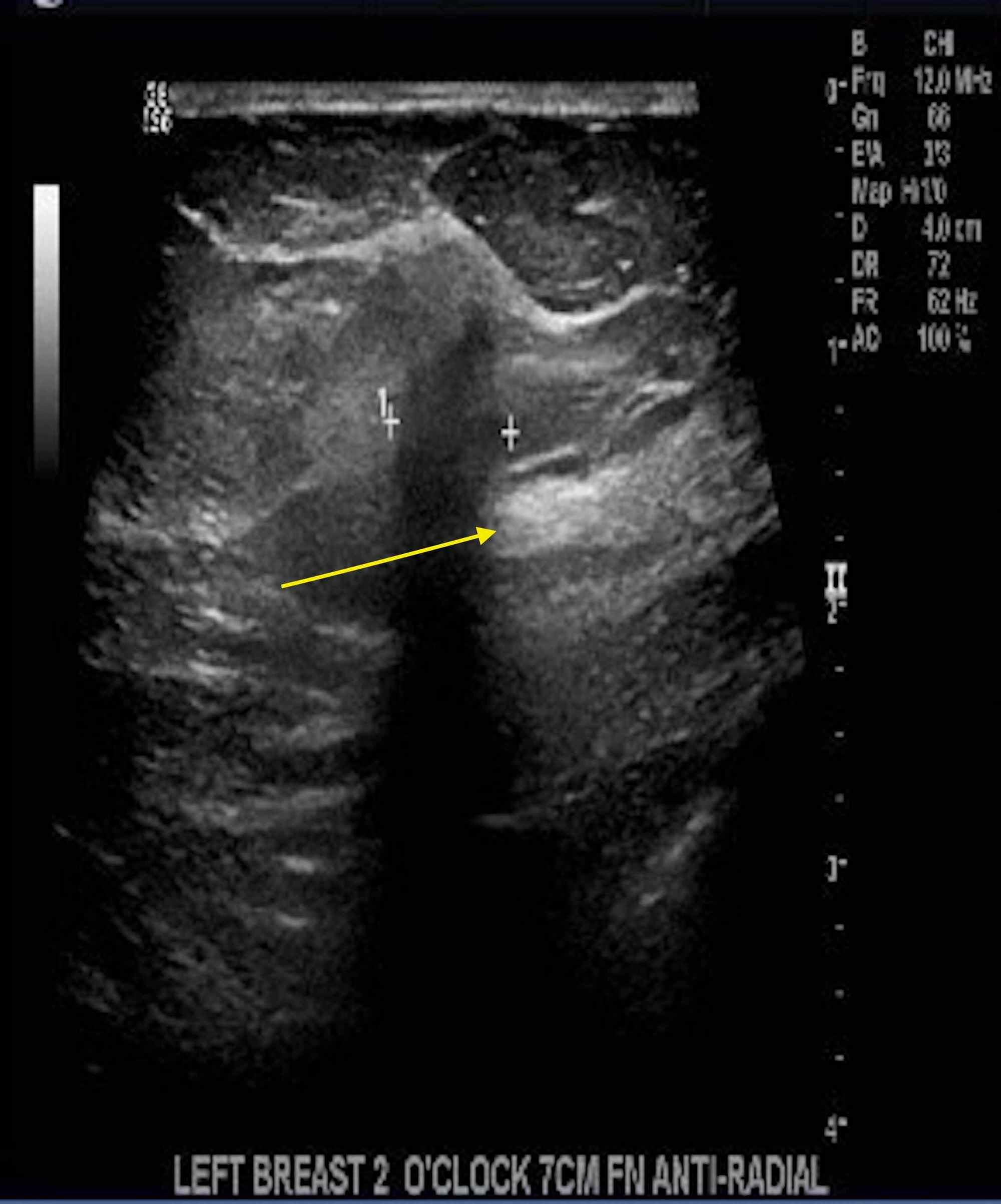
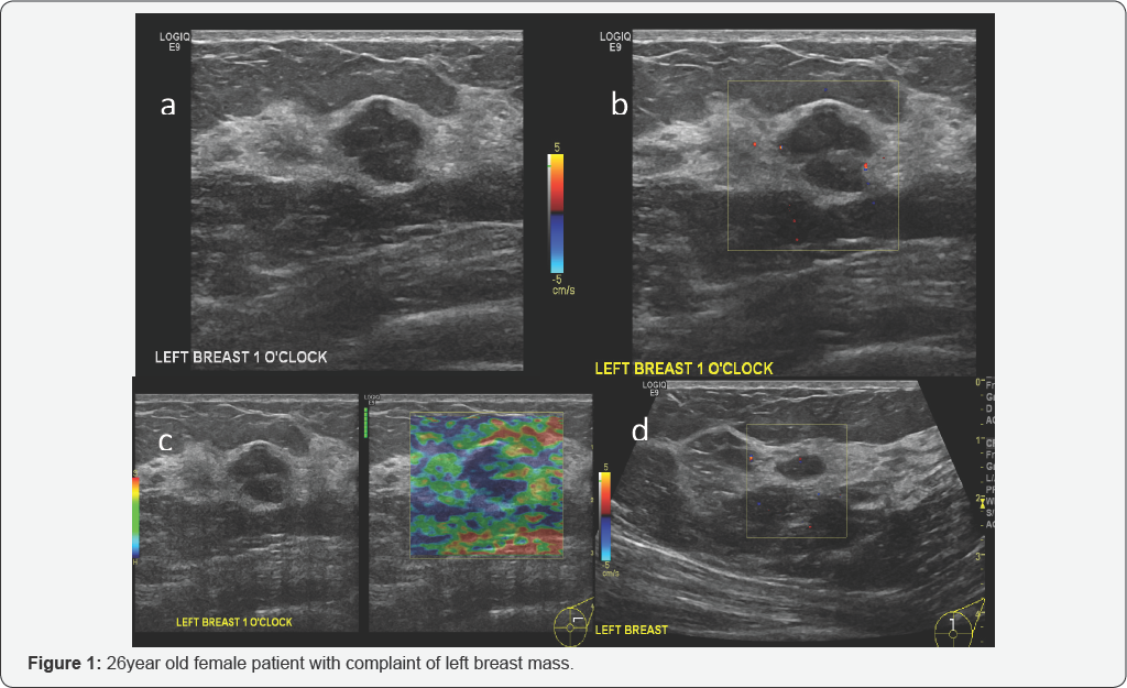
Case Discussion MACROSCOPIC DESCRIPTION "Left breast 1 o'clock, 7cm FN" Four cores 30mm in total length A1 MICROSCOPIC DESCRIPTION Sections show multiple cores of breast parenchyma which shows an invasive carcinoma with morphological features in keeping with a BRE grade 2 tumor DIAGNOSIS Left breast 1 o'clock 7cm FN Invasive carcinoma NST The positive predictive value on a screening examination for masses and calcifications is similar and is slightly lower for developing asymmetry and least for focal asymmetry Although architectural distortion is the least common of the four frequent signs of breast cancer, its reported positive predictive value for breast cancer on a screening examination (102 %) is Palpation of Benign Breast Masses In contrast to breast cancer tumors, benign lumps are often squishy or feel like a soft rubber ball with welldefined margins They're often easy to move around (mobile) and may be tender 4 Breast infections can cause redness and swelling
Gender Female Testing 3D Mamography Both Breast Case Cervical Cancer (Surgery Done) My mother had cervical cancer and the surgery has been done After surgery we have done the 3D Mamography Both Breast test and in findings i see below HRUS Evidence of 3x3 mm well defined anechocic cyst is seen at 10 o'clock position in left breast BIRADSII Can anyone explain theAn initial mammogram showed a hypoechoic solidappearing mass at the 10o'clock position which was 22×2×17 cm with irregular lobulated borders concerning for malignancy in her right breast Family history was negative for breast cancer, but positive for prostate cancer on Magnetic resonance imaging (MRI), using a T1weighted image, showed enhancement of the lesion (Fig 3a, b)The left mass was located at the 10 o'clock position in the left breast, and the right mass was located at the 12 o'clock position in the right breast The implants were located under the pectoralis major muscle


Individualized Treatment Analysis Of Breast Cancer With Chronic Renal Ott
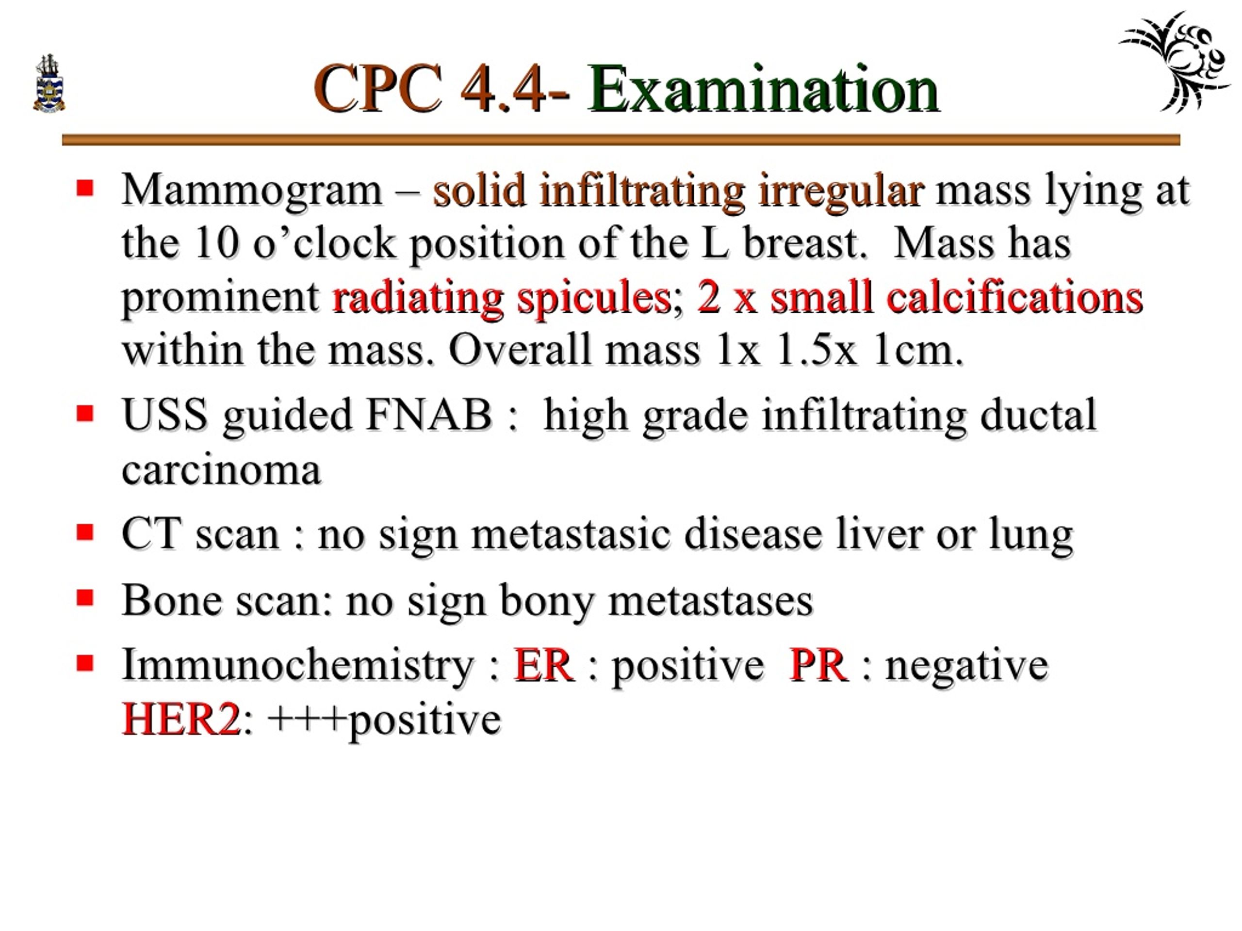


Ppt Pathology Of Breast Disorders Powerpoint Presentation Free Download Id
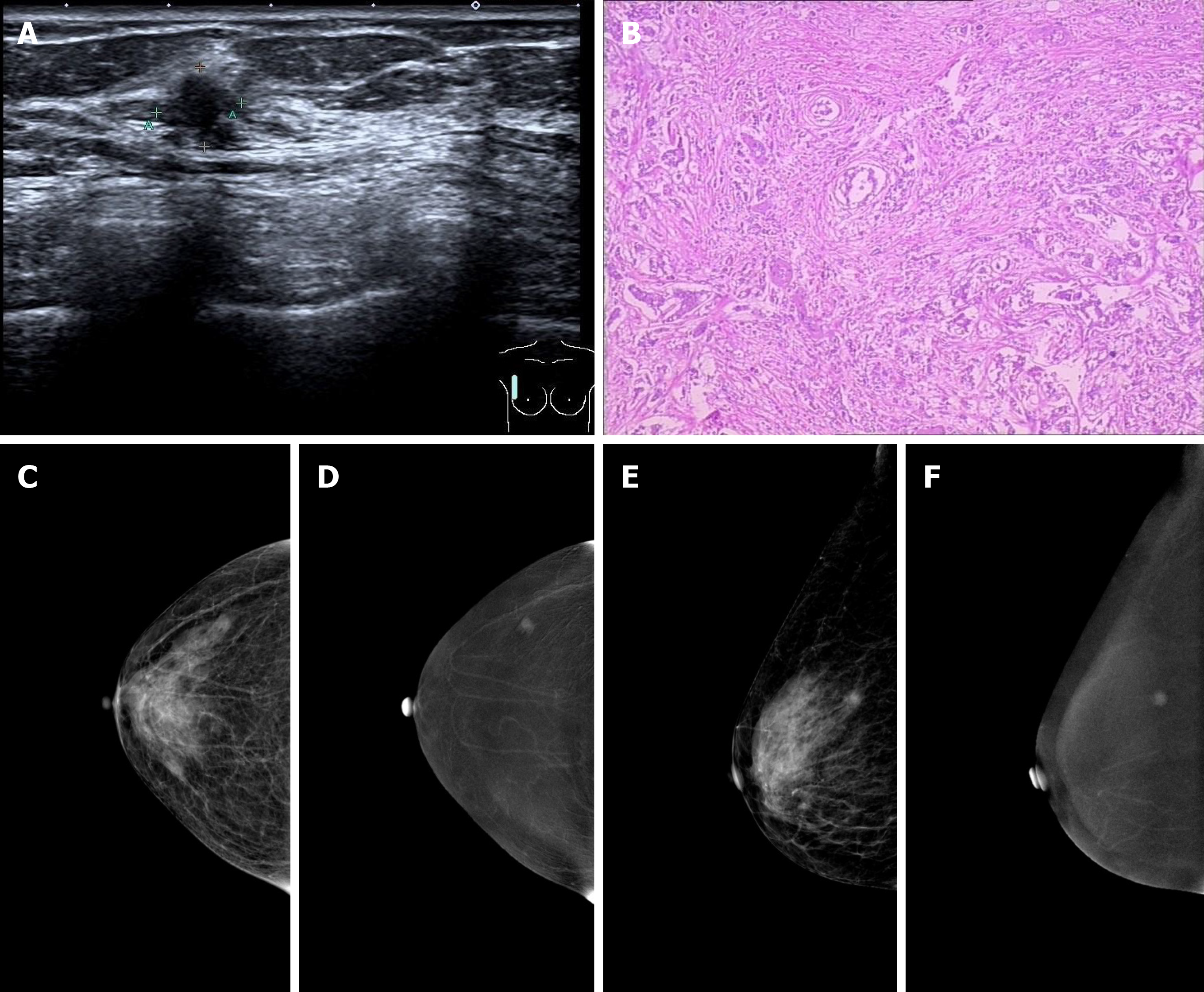
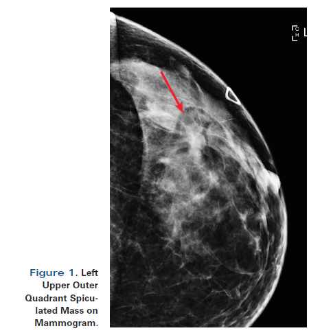
Tumor size is an important factor in breast cancer staging, and it can affect a person's treatment options and outlook Tumors are likely to be smaller when doctors detect them early, which can Breast cancer, ultrasonography Mediolateral oblique digital mammogram of the right breast in a 66yearold woman with a new, opaque, irregular mass approximately 1 cm in diameter The mass has spiculated margins in the middle third of the right breast at the 10o'clock position Breast ultrasonography revealed a hyperechoic view of the entire skin of the right breast and a 36mm irregular mass at the 9 o'clock position (Fig 2) A T2weighted magnetic resonance imaging (MRI) examination showed a highsignal mass at the same area, and enlargement of the right axillary lymph nodes (Fig 3 a, b)
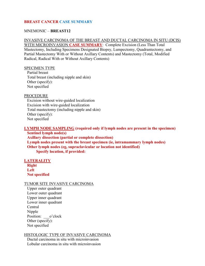


Breast Invasive Carcinoma


3
The largest cyst in the (L) breast is alos at 3 o'clock position A cystic lesion at 4 o'clock position has an internal septation within There is on benign appearing solid lesion at 10 o'clock position in the (R) breast measuring from the nipple This is stable since the previous study In the (L) breast, there is a solid lesion at 4 o'clock position, 5 cm from the nipple3 o'clock position (right breast) is towards the middle of your chest3 o'clock position (left breast ) is towards your armpitHope I've been helpfulRegards Comment japdip Breast cancer is not an inevitability From what you eat and drink to how much you exercise, learn what you can do to slash your risk The position of the tumor in the breast may be described as the positions on a clock Disrupted expression of circadian genes can alter breast biology and may promote cancer There is a single tumor located at the 12 3 6 or 9 oclock position on the breast code the primary site to c509 when there are multiple tumors two or more in at least two quadrants of the breast


Methylene Blue Dye Related Changes In The Breast After Sentinel Lymph Node Localization Kang 11 Journal Of Ultrasound In Medicine Wiley Online Library


1
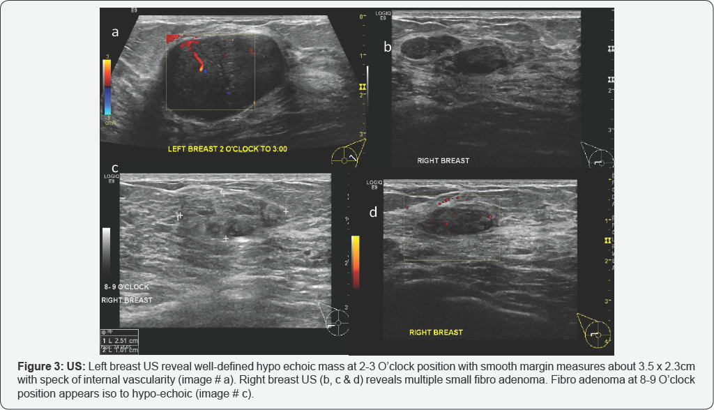
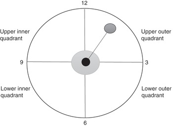
The least frequent location of breast cancer is the lower inner quadrant Another way to describe the location of frequency is to look at the breast like a clock Breast cancer is more frequent between 12 and 3 of the left breast and between 12 and 9 in the right breast The distribution of breast cancer is related to the tissue volume of the breastO'Clock Positions and Codes Quadrants of Breasts 2 11 12 1 1 10 9 8 7 7 6 5 5 4 3 2 11 12 10 6 4 RIGHT BREAST LEF T BREAST UOQUIQ LOQ LIQ LIQ LOQ C502 C504 C502 C504 C505 C503 C503 C500 C501 SEER Program Coding and Staging Manual 07 C606 SiteSpecific Coding Modules Appendix CThree hypoechoic nodules at 3 o'clock position measures about 24 x 10 mm, 10 x 3 mm and 7 x 4 mm (d)with no internal vascularity in Doppler exam (not shown) (Figure 2) ImpressionCase 2 Abovedescribed finding representing bilateral breast fibro adenomas BIRADS 3
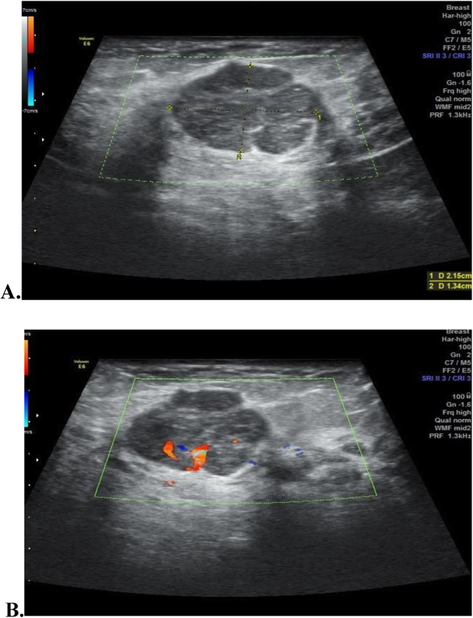


Ultrasound Lexicon In Diagnosis And Management Of Breast Fibroadenoma When To Follow Up And When To Biopsy Egyptian Journal Of Radiology And Nuclear Medicine Full Text
/know-your-breast-tumor-size-4114640-FINAL-f17fb19bf9214d20937d07bd41524ac7.png)


Breast Tumor Size And Staging
Review of a mammography study performed 2 years previously demonstrated no evidence of theThe right breast was normal to palpation Mammography revealed a solid, smooth nodule in the upper internal quadrant of the left breast (Fig 1) Ultrasound demonstrated an oval mass in the 10o'clock position of the left breast measuring 15×10 mm (Fig 2)In the diagram below, the nipple is in the center of the clock for both breasts The outer left breast is at 3 o'clock and the outer right breast is at 9 o'clock In the left breast the upper outer quadrant is between 12 and 3 o'clock
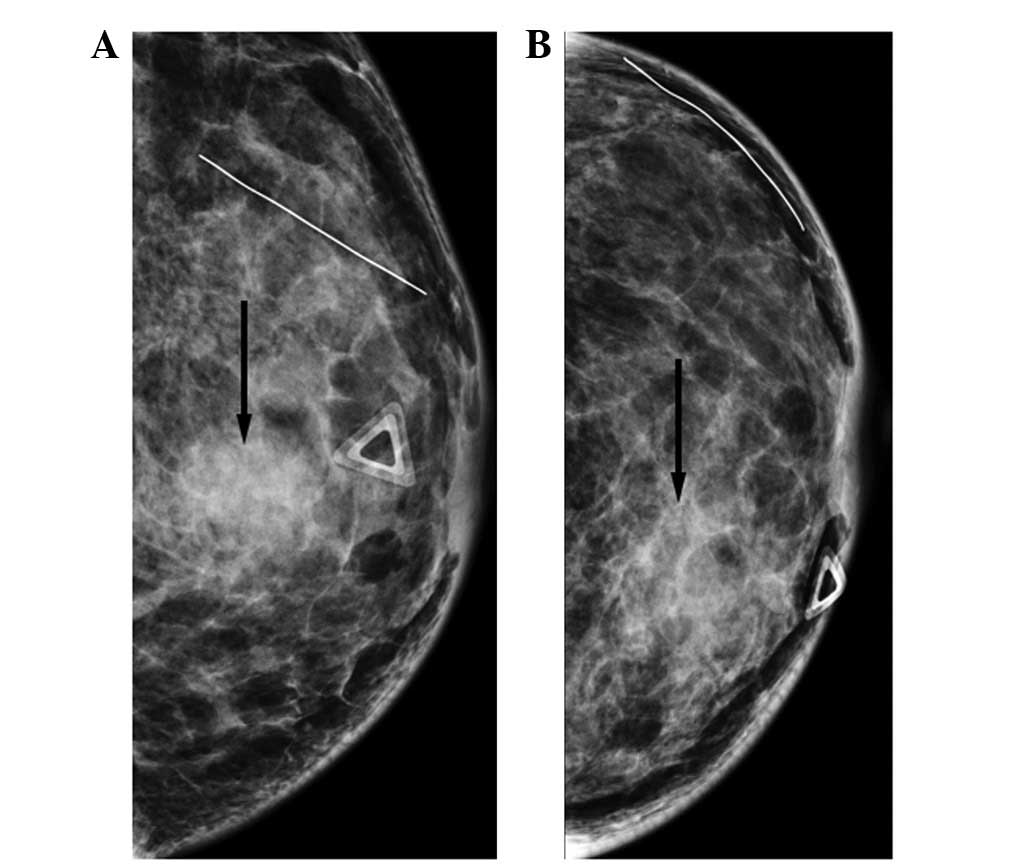


Breast Metastasis From Nasopharyngeal Carcinoma A Case Report And Review Of The Literature


Recent Comprehensive Review On The Role Of Ultrasound In Breast Cancer Management Leaders In Pharmaceutical Business Intelligence Lpbi Group
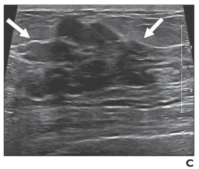
A 50yearold woman was found on screening mammography to have suspicious pleomorphic microcalcifications in the 12o'clock position of the right breast Biopsy was performed with stereotactic technique, confirming intermediategrade ductal carcinoma in situ (DCIS) ( Figure 2 )There were nonmass lesions of about 22 cm wide with internal calcification extending to the subareolar area at the 2–3 o'clock position of the right breast in the breast ultrasound Additionally, there was a 06cm low suspicious finding at the 10 o'clock position of the right breast in the breast ultrasound examinationABUS detected a right breast irregular hypoechoic mass with angular margins and posterior acoustic shadowing at the 1030 o'clock position, measuring 15 x 16 x 11 cm, 7 cm from the nipple Dedicated handheld ultrasound confirmed the mass A core biopsy was performed and detected invasive moderately differentiated ductal carcinoma


1


The Radiology Assistant Bi Rads For Mammography And Ultrasound 13
Corresponds to an irregular mass lesion with indistinct margins at the 10 o'clock position, 30 mm from the nipple Combined imaging category 5 Malignant Architectural distortion and stellate lesion Architectural distortion represents lesions seen on mammography that cause distortion of the normal architecture of the breast parenchyma The margin is irregular Another three hypoechoic nodules noted at 8 and 10 o'clock position of right breast and behind the nipple (05 x 07 cm, 05 x 05 cm and 17 x 18 cm) A right axillary node is also seen, 09 x 08 cm Impression Findings in keeping with Ca breast(a) Targeted US image obtained at the 6o'clock to 10o'clock position in the right breast for initial evaluation shows a large complex mass (arrowhead) with solid and cystic components and internal vascularity The mass was believed to represent surrounding inflammation and/or granulation tissue associated with abscess formation and was classified as a BIRADS category 3


Fcds Med Miami Edu Downloads Naaccr Webinars Breast Breast final Pdf


Casereports Bmj Com Content 15 r 14 67 Full Pdf
Introduction Inflammatory breast cancer (IBC) is a rare subtype of breast cancer that accounts for 2%–5% of all breast cancers It has a highly virulent course with a low 5year survival rate of 25%–50% ()Trimodality treatment that includes preoperative chemotherapy, mastectomy, and radiation therapy is the therapeutic mainstay and has been shown to improve prognosis (2–4)At the exam, there is a fistula in 11 o'clock position the right breast from which came out pus, associated with a palpable mass She was initially treated by ultrasoundguide drainage of the abscess, associated with a correct antibiotic correlated with the sensitivity of the germ (staphylococcus aureus) isolated by bacteriology at the clinic without any improvement Breast cancer, ultrasonography The patient in Images 2628 also had a 7mmdiameter cyst at the 10o'clock position in the right breast


Chapter 3 Lange Improved Flashcards Quizlet
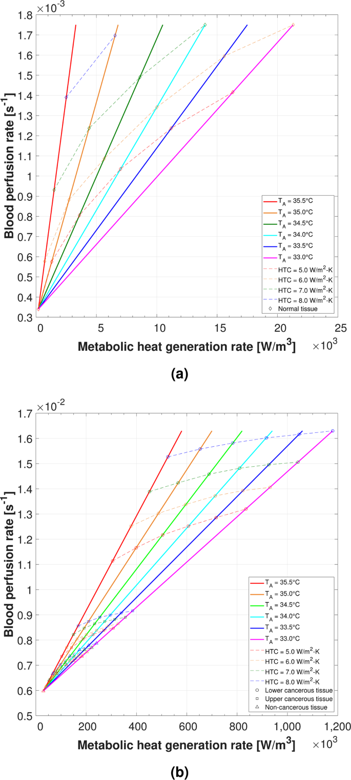


Determining The Thermal Characteristics Of Breast Cancer Based On High Resolution Infrared Imaging 3d Breast Scans And Magnetic Resonance Imaging Scientific Reports
Breast Cancer ICD10 Code Reference Sheet FEMALE Right C Malignant neoplasm of nipple and areola, right female breast C Malignant neoplasm of central portion, right female breast C Malignant neoplasm of upperinner quadrant, right female breastA 42yearold woman who was diagnosed at her local hospital with invasive breast cancer at the 10 o'clock position of her right breast by core needle biopsy was referred to our department in October 19 Physical examination revealed a welldefined palpable mass in the upper outer quadrant of the right breast In the left breast there are two adjacent oval solid lesions measuring 5 and 65mm in diameter present in the 2 o'clock position In the 7 o'clock position there is a larger oval lesion measuring 10 x 5mm The appearance of each of the three lesions within the left breast is consistent, but not diagnostic of fibroadenoma


Cureus Invasive Lobular Breast Carcinoma Can Be A Challenging Diagnosis Without The Use Of Tumor Markers


Invasive Micropapillary Carcinoma Of The Breast Radiology Case Radiopaedia Org
In the Introduction, Dr Ellis wrote, "Due to these positional differences, a lesion reported at the 10o'clock position of the breast with mammography may be much closer to the 8o'clock position with sonography" Although confusion between different quadrants may occur for the lesions near the 3, 6, 9, or 12o'clock positions, the 10o'clock and 8o'clock positions are in completelyOne study analyzed almost 2,000 breast tumor samples and determined that 10 separate breast cancer subtypes could be identified, based on clusters of genes that were either expressed or not, in particular patterns 7 Many of these gene clusters alsoArea of irregular heterogeneous breast tissue in right upper, outer quadrant 10o'clock position (arrow) Core needle biopsy showed invasive lobular cancer at histopathology, consistent with multicentric disease (d) Extendedfieldofview grayscale ultrasound shows two separate masses in right 7 and 10o'clock positions, 5 cm apart


Cureus Diagnosis Prognosis And Management Of Breast Cancer In An 81 Year Old Male Patient


Breast Cancer Signs Symptoms And Understanding An Imaging Report Saint John S Cancer Institute
On 14 March 14, WF had an ultrasound of her breasts "There is a 17 mm x 96 mm lesion at 2 o'clock position of left breast, 4 cm from the nipple" A FNAC (Fine needle aspiration cytology) done in a Taiping private hospital showed "benign breast• there is a single tumor located at the 12, 3, 6, or 9 o'clock position on the breast Code the primary site to C509 when there are multiple tumors (two or more) in at least two quadrants of the breast Laterality Laterality must be coded for all subsites At ultrasonography, a second illdefined, hypoechoic, heterogeneous mass was identified at the 10 o'clock position in the right breast, measuring 1 mm × 10 mm × 1 mm The secondary lesion was retrospectively visualized on the screening mammogram as well (Figs 1c, d) Both masses underwent core biopsy by ultrasound guidance


Coding Breast Mass Becomes More Specific For Coders


Diagnosis And Management Of Benign Breast Disease Glowm
10 o'clock position on mammogram () Recent clinical studies Diagnosis Cavernous hemangioma of the breast mammographic and sonographic findings and followup in a patient receiving hormonereplacement therapy Mesurolle B,Wexler M,Halwani F,Aldis A,Veksler A,Kao EJ Clin Ultrasound03 Oct;31(8)4306doi /jcu Clock face description of breast lesion locations The clock face location of breast findings is described by imaging a clock on both the left and the right breast as the woman faces the examiner Note that the outer portion of the breast on the right is at the 9o'clock position and the outer portion on the left is at the 3o'clock position A 58yearold woman presented with a right breast lump at the 6 o'clock position Mammography showed a 3cm illdefined mass with irregular margins and associated architectural distortion abutting the chest wall in the lower outer quadrant of the right breast (Fig 3 ( 7K ));


Diagnostic Breast Imaging Clinical Breast Imaging A Patient Focused Teaching File 1st Edition


Antr510 027 Anatomy Of The Breast Msu Mediaspace
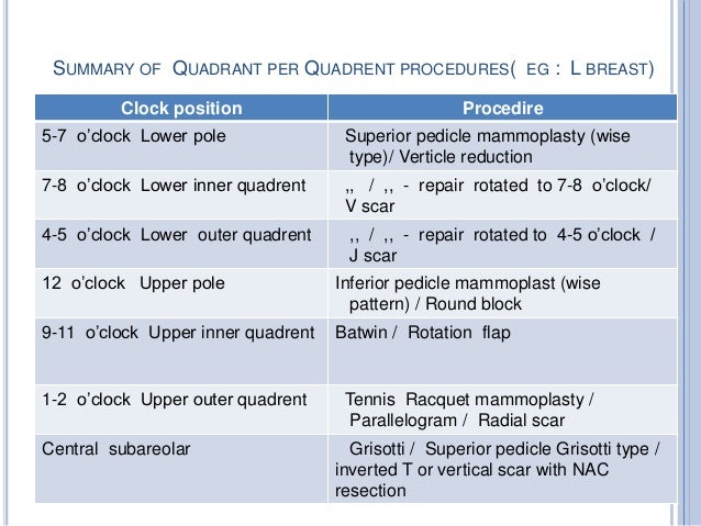
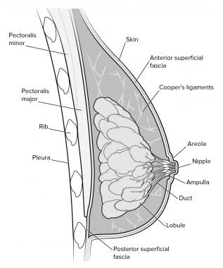
In the right breast, the nerve generally enters the posterolateral surface of the breast at the 8 o'clock position and traverses the gland to enter the right areola along the 7 o'clock axis Avoiding the trajectory of these nerves, particularly when performing circumareolar incisions and advancement flaps, will preserve nippleareolar complex sensation and improve the quality of life ICD 10 AM Edition Eleventh Edition Query Number 3598 In the Eleventh Edition Education slides Neoplasms, there is a diagram with clock positions with an example of documentation of a breast lesion at 6 o'clock coded to C508 Overlapping lesion of breast VICC #3107 Breast Quadrants and neoplasm codes advises to use a diagram as a reference to assist


Primary Breast Cancer Patient With Poliomyelitis A Case Report


Bilateral Breast Cancer Different In More Ways Than One


Breast Coding Guidelines Surveillance Epidemiology And Flip Ebook Pages 1 4 Anyflip Anyflip


The Radiology Assistant Bi Rads For Mammography And Ultrasound 13


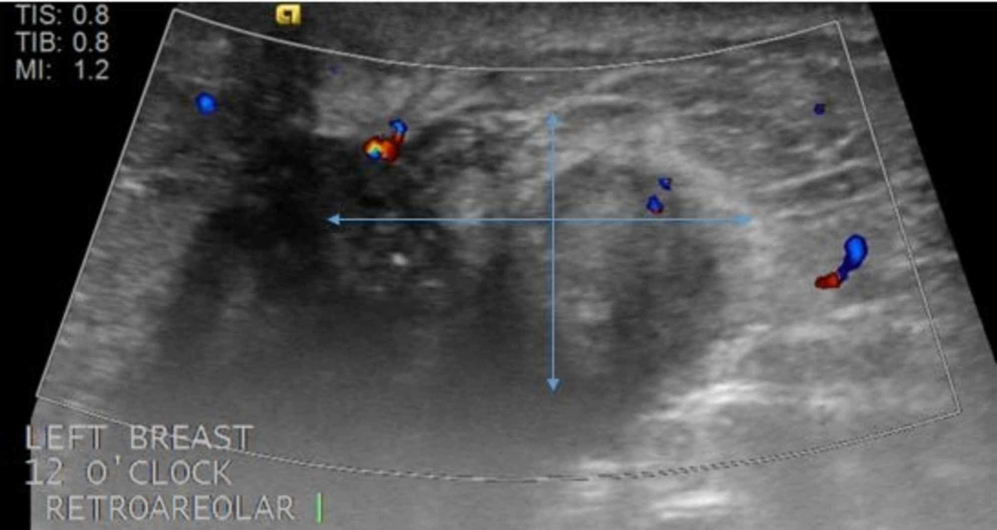
Ultrasonography At The 12 O Clock Position Of The Left Breast Revealed Download Scientific Diagram


Breast Cancer


Cancers Free Full Text Primary Neuroendocrine Neoplasms Of The Breast Case Series And Literature Review Html


Undiagnosed Breast Cancer Features At Supplemental Screening Us Radiology


Epos


Breast Reconstruction
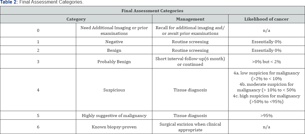


Cancer Therapy And Oncology International Journal Ctoij


Hypothetical Chief Complaint Ppt Download


Breast Cancer Ultrasonography Practice Essentials Role Of Ultrasonography In Screening Breast Imaging Reporting And Data System


How To Self Examine Your Breast You Me Medicine
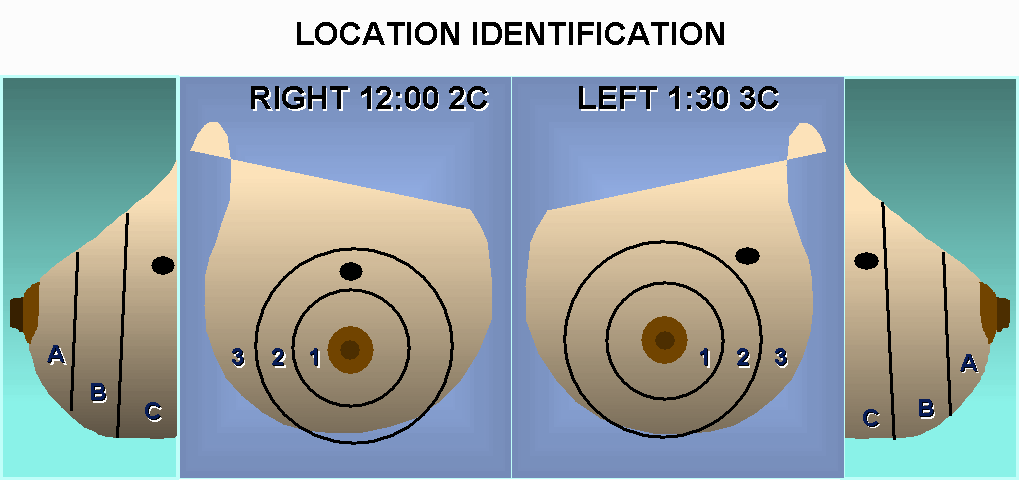


Sonography Of The Breast


Icd 10 Cm Coding Overlapping Breast Quadrants Coffee With A Medical Coding Auditor


Mammography In Breast Cancer Background X Ray Mammography Ultrasound


Usefulness Of 3d Surgical Guides In Breast Conserving Surgery After Neoadjuvant Treatment Scientific Reports


Circumscribed Masses Medium Or High Density Masses Radiology Key


Breast Mri For Diagnosis And Staging Of Breast Cancer Springerlink


Early Diagnosis And Treatment Of Cancer By Juan Jaramillo Issuu


Journal Of Lancaster General Health Depart3 V1i2


Seer Cancer Gov Manuals 21 Appendixc Coding Guidelines Breast 21 Pdf


Breast Mass Radiology Key


A 55 Year Old Woman With New Triple Negative Breast Mass Less Than 2 Cm On Both Mammogram And Ultrasound


Post Mastectomy Radiation After Immediate Reconstruction For Multifocal Early Stage Breast Cancer To Irradiate Or Not International Journal Of Radiation Oncology Biology Physics


Www Birpublications Org Doi Pdf 10 1259 Bjr


Lactational Breast Changes Lobular Hyperplasia Mimicking Masses How Can We Differentiate From True Pathological Masses


Contrast Enhanced Dedicated Breast Ct Detection Of Invasive Breast Cancer Preceding Mammographic Diagnosis Sciencedirect


26 Year Old With Palpable Mass Near The 12 00 O Clock Position Of The Download Scientific Diagram


Print Anatomy Physiology And Pathology Of The Breast Mammography Flashcards Easy Notecards


Diagnostic Breast Imaging Clinical Breast Imaging A Patient Focused Teaching File 1st Edition


Factors Affecting Visualization Rates Of Internal Mammary Sentinel Nodes During Lymphoscintigraphy Journal Of Nuclear Medicine


O Clock Position Of A Lesion On A Right Breast As Identified By Download Scientific Diagram


Fcds Med Miami Edu Downloads Naaccr Webinars Breast Breast final Pdf


Usefulness Of Postoperative Surveillance Mr For Women After Breast Conservation Therapy Focusing On Mr Features Of Early And Late Recurrent Breast Cancer


Cancer Therapy And Oncology International Journal Ctoij


National Program Of Cancer Registries Education And Training
/know-your-breast-tumor-size-4114640-FINAL-f17fb19bf9214d20937d07bd41524ac7.png)


Breast Tumor Size And Staging


Contrast Enhanced Dedicated Breast Ct Detection Of Invasive Breast Cancer Preceding Mammographic Diagnosis Sciencedirect


Experts To Consider All Aspects Of Breast Ultrasound European Society Of Radiology


Nonvisualization Of Sentinel Node By Lymphoscintigraphy In Advanced Breast Cancer Topic Of Research Paper In Clinical Medicine Download Scholarly Article Pdf And Read For Free On Cyberleninka Open Science Hub
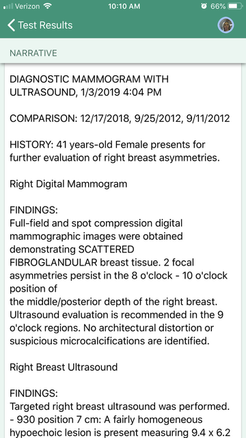


Breast Cancer Topic Interpreting Your Report


Ispub Com Ijanp 12 1 2947


Breast Cancer Topic If You Are Waiting Please Let Me To Share My Experience


Nucleus Iaea Org Hhw Radiology Clinical Applications Specialities And Organ Systems Breast Imaging Guidelines And Literature Suggested Articles Reading A Mammogram Pdf
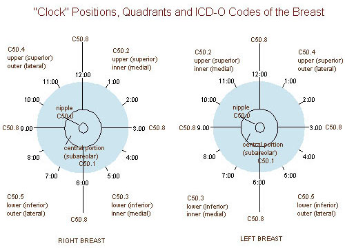


Seer Training Quadrants Of The Breast


Breast Cancer Lumps Where Are They Usually Found


Http Static pc Com A3c7c3fe 6fa1 4d67 8534 A3c9c15fa0 C7b39f96 0935 4e94 84b8 C4a6d9d86abd 3739cb 8b1c 4e15 6b E6192eec5078 Pdf


Breast Cancer Topic Quadrants Of Breast Cancer Survey Also


Nucleus Iaea Org Hhw Radiology Clinical Applications Specialities And Organ Systems Breast Imaging Guidelines And Literature Suggested Articles Reading A Mammogram Pdf


Www Biobran Org Uploads Case Study 3a1d32a1d152e7f1b1c5818d322b81ac Pdf
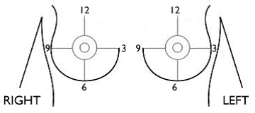


Breast Cancer Signs Symptoms And Understanding An Imaging Report Saint John S Cancer Institute
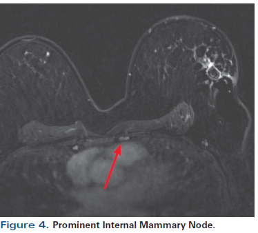


A 55 Year Old Woman With New Triple Negative Breast Mass Less Than 2 Cm On Both Mammogram And Ultrasound


2


Www Archbreastcancer Com Index Php Abc Article Download 238 373


Office Care Of Breast Disorders Chapter 34 Office Care Of Women


Ultrasonography At The 12 O Clock Position Of The Left Breast Revealed Download Scientific Diagram


Coding Breast Mass Becomes More Specific For Coders


Spie Org Samples Pm211 Pdf


Breast Mri Identified Lesions During Neoadjuvant Therapy Are Largely Benign


Coding Breast Mass Becomes More Specific For Coders


2


Preoperative Tumor Size Measurement In Breast Cancer Patients Which Threshold Is Appropriate On Computer Aided Detection For Breast Mri Cancer Imaging Full Text


Osa Three Dimensional In Vivo Fluorescence Diffuse Optical Tomography Of Breast Cancer In Humans


Gallery Formats Parvizbagloo


Spie Org Samples Pm211 Pdf


Mammography What Are The Common Clinical Problems Presenting As Breast Mass Common Breast Masses Are Cyst Fibroadenoma Cancer What Is The Utility Of The Following Imaging Studies In The Evaluation Of Breast Mass Mammography Ultrasound Ct


Diagnosis And Management Of Benign Breast Disease Glowm


Lange Q A Chapter 3 Anatomy Physiology And Pathology Of The Breast Flashcards Quizlet


Finding Early Invasive Breast Cancers A Practical Approach Radiology


Figure 2 From Efficacy Of Pectoral Nerve Block Type Ii For Breast Conserving Surgery And Sentinel Lymph Node Biopsy A Prospective Randomized Controlled Study Semantic Scholar


Breast Diseases Obgyn Key


Http Www Kumc Edu Kcr Newsletters Kcr Newsletter October16 Pdf


Http Journals Sagepub Com Doi Pdf 10 1177


Fcds Med Miami Edu Downloads Teleconferences 11 11 Breast Fcds Required 3 Per Page With Note Space Pdf


Cancer Therapy And Oncology International Journal Ctoij


Icd 10 Code For Breast Cancer At 12 00


The Role Of Ultrasound In Breast Imaging Document Gale Onefile Health And Medicine


Q Tbn And9gcth39qrs02wrthtpk Uisqk0p0rnefiz Yi0mmwn2cv4eblwxg Usqp Cau


0 件のコメント:
コメントを投稿